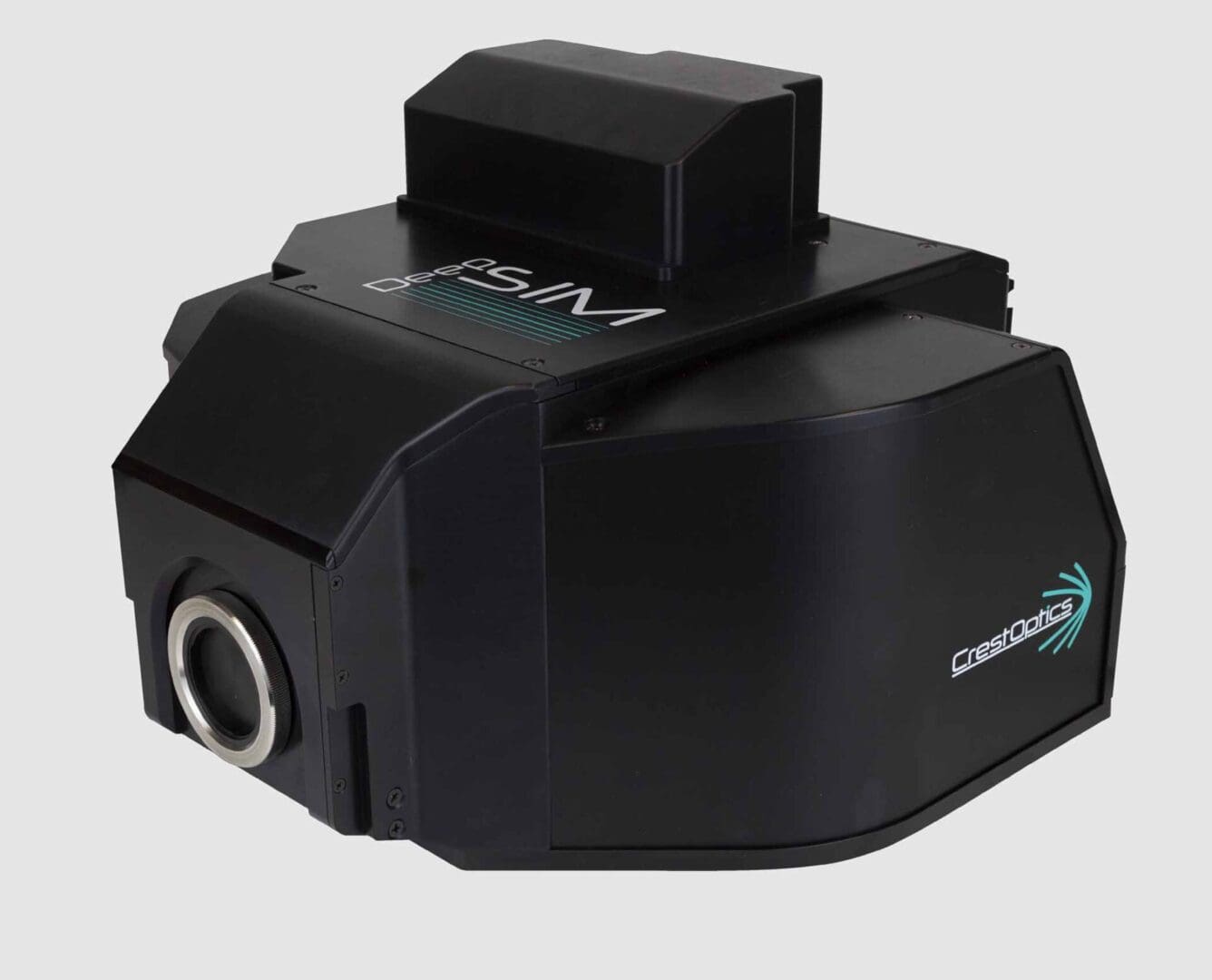
CrestOptics DeepSIM Imager
- Available in two versions: Stand alone or with CrestOptics X-Light V3 Spinning Disk.
- FOV:1024x1024pixel (66×66 µm 100X | 333×333µm 20X).
- Resolution: Lateral Resolution (FWHM): ~100 nm (100X NA 1.45) Axial Resolution (FWHM): ~300 nm (100X NA 1.45).
- Software: µManager /VisiView® / NIS Elements.
- Laser spectral range: Excitation: 400-750 nm; emission: 400-850 nm.
- Camera compatibility: Any triggerable camera having 6.5 µm pixel size.
Description
Based on a multi-spot structured illumination system, the DeepSIM is a reliable, simple to use and affordable solution to study cellular structures up to an XY resolution of ~100nm.
Super-resolved optical sectioning, with Z resolution up to ~ 300nm, can be obtained using both high (60X – 100X) and low magnification (20x – 40x) objectives to expand the range of applications to include complex 3D models such as tissues, organoids, spheroids and small organisms. The DeepSIM is designed to work with samples of thicknesses comparable to those used in confocal microscopy, giving super-resolved data over 50µm Z in depth in non-clarified samples. DeepSIM enables the effortless study of live-cell dynamics through a temporal resolution greater than 10fps (1024×1024 px FOV), allowing biological changes to be tracked at cellular and subcellular levels. DeepSIM is designed to be a truly enabling technology where performance is combined with the flexibility of a system available either as a super-resolution module combined with CrestOptics X-Light V3 Spinning Disk or as stand-alone equipment. The DeepSIM constitutes the seamless evolution of any diffraction-limited microscopy approach. It is as easy to use as a widefield system and reaches the same Z-depth penetration of a confocal microscope, enabling scientists to access super-resolved data through conventionally prepared, deep, thick specimens, even in challenging sampling conditions. The 2D Lattice SIM technology behind DeepSIM enhances the illumination efficiency and homogeneity with minimal phototoxicity in live cell imaging experiments. Besides, the 2D Lattice SIM Illumination delivers a better contrast that significantly improves in-depth Z-acquisitions and the image reconstruction robustness. More importantly, the user can choose among 3 Lattice SIM Illumination Patterns for approaching specimens of increasing thickness and morphological complexity. The well-matched sampling conditions significantly mitigate the risk of artifacts in image reconstruction. Featuring the structured lattice illumination with the computationally super-resolved data reconstruction, DeepSIM improves the spatial resolution in all three dimensions reaching 100 nm laterally and 300 nm axially. Deep super-resolved data across conventionally prepared thick samples. The DeepSIM is designed to work with samples of thicknesses comparable to those used with Confocal microscopy, giving super-resolved data over 50 μm Z-depth. Z-stacks take full advantage of the entire objective working distance and meaningful data can be obtained from a native heterogeneous and intricate sample, providing researchers with a complete understanding of its structure and composition. Multiplexed imaging is an emerging way to gather information from multiple cellular markers simultaneously. DeepSIM performs optimally with the standard staining markers for confocal microscopy. It is the only SIM technology covering the full spectrum from 405 nm to NIR 750 nm excitation wavelength. DeepSIM offers the maximum flexibility in fluorophore choice and reduces the spectral overlap in multicolor experiments. The DeepSIM was conceived to maintain high standards of performance over a large set of objectives from Low Magnification 20x up to High Magnification 100x. The compatibility with dry lenses and any immersion media expands the range of suitable applications over a large cohort of sample and support types. DeepSIM offers the best trade-off between FOV and resolution to catch fine details in extra-large objects. The DeepSIM resolving power is preserved across a wide spatial scale, including 3D organoids, tissue slices, and small animal models (Zebrafish, C. elegans). The extended flexibility suggests its use with Spinning Disk technology in a correlative bioimaging approach. DeepSIM is designed to be a truly enabling technology where performance is combined with the flexibility of a modular, expandable system. DeepSIM can seamlessly upgrade any existing frame with a camera port, be it upright or inverted, thus leading to a significant cost-saving setup.

Reviews
There are no reviews yet.